
Microstructural characterization
|
|
|
|
| Electron microscopy | X-ray diffraction | Optical microscopy |
|
|
|
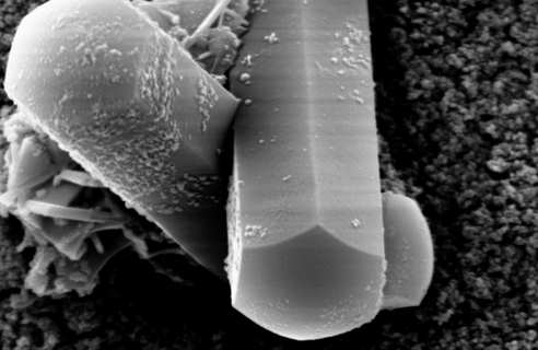
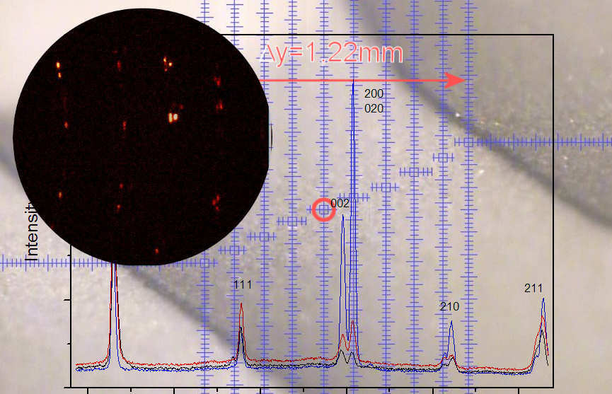
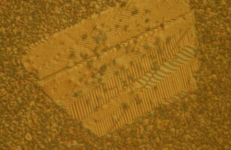
The macroscopic properties of ceramic materials are determined by the microstructure that forms during sintering to create the finished material. Microstructural characterization is therefore one of the central cross-sectional tasks, both for process technology and for the evaluation of properties and service life. Optical microscopy, imaging and analytical electron microscopy and X-ray fine structure analysis are used as classic characterization methods. Building on this, innovative characterization methods are developed in collaboration with other working groups, such as X-ray diffraction contrast tomography for 3D visualization of ceramic material structures.
Research in the working group focuses, among other things, on densification and grain growth during sintering. The aim here is to develop an understanding of the processes during the movement of grain boundaries. The methodological approach ranges from the atomic scale locally at the grain boundaries to the movement behavior of the grain boundaries and the classic parameters such as average grain size and distribution width.